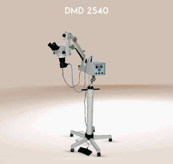
An Ophthalmic Operating Microscope (OOM) is a specialized Ophthalmic Equipment that is designed for use in ophthalmic surgery. It plays a crucial role in providing high-resolution, stereoscopic images of the eye during surgical procedures, enabling surgeons to perform delicate and precise interventions. It helps to perform toughest surgery by providing high-resolution images and details.
This sophisticated microscope is equipped with features tailored to meet the unique requirements of eye surgery, offering high magnification, excellent depth perception, and illumination.
An operating or surgical microscope is ophthalmic equipment that provides the surgeon with a stereoscopic, high-quality magnified and illuminated image of the small structures in the surgical area. This is essential for a detailed examination of different parts of the eye, such as the cornea, lens, retina, and other structures. This microscope is designed with fine-focus mechanisms to help surgeons achieve sharp and clear images.
These microscopes can often be mounted on floor or celling, which allows surgeons to position the microscope comfortably for different procedures and also helps to make movement easier.
It has objective lens with eyepiece of 45º inclined. It has features like magnification, illumination systems, binoculars and stereoscopic vision, Adjustability and manoeuvrability and camera systems. These features make it the crucial and important microscope in the surgery of eye providing surgeons with the tools needed to perform intricate procedures with a high level of precision.
What is the use of operating microscope?
An Ophthalmic microscope intended for use in a surgical setting—particularly necessary for microsurgery—is called a surgical microscope, sometimes referred to as an operating microscope.
What is the working distance of the ophthalmic microscope?
Ophthalmic surgery, objective focal lengths of 150 mm, 175 mm, and 200 mm are frequently utilized for complex surgeries in the back area.
What is the ophthalmic microscope?
An ophthalmic microscope is a specialized microscope used by ophthalmologists for examining the eye's anterior segment and posterior segment. It provides a magnified, three-dimensional view of the eye's structures, including the cornea, iris, lens, and anterior chamber.
The ophthalmic microscope has revolutionized the field of eye surgery, providing surgeons with the tools needed to perform intricate procedures with a high level of precision. As technology continues to advance, we can expect further refinements and innovations in ophthalmic microscope design, contributing to improved patient outcomes in the field of ophthalmology.